
- 実験動物の対物攻撃行動を機械で計る・Calculating aggressive behavior towards inanimate objects using a machine
- 精神疾患モデルマウスの攻撃行動を調べる・Analysis of aggressiveness in psychiatric model mice
- 光くしゃみ反射
Photic Sneeze Reflex - 特定の脳神経細胞に薬を届ける
- 脳の局所血流調節を司る細胞
- 頭部・顔面・口腔領域の血流調節
- 血管調節細胞と分泌調節細胞の形態差
- 副毛様体神経節の発見
- 副毛様体神経節の解剖学
- 今も続くダイオキシン汚染
- ダイオキシンの次世代への脳影響
- ダイオキシンによる前脳の異常
- ダイオキシンによる脳エンケファリン異常
- ダイオキシン摂取による拒食症
- 一酸化窒素が摂食とエネルギーバランスに関係する?
私たち研究グループの研究・Research of our group
光くしゃみ反射
Photic Sneeze Reflex
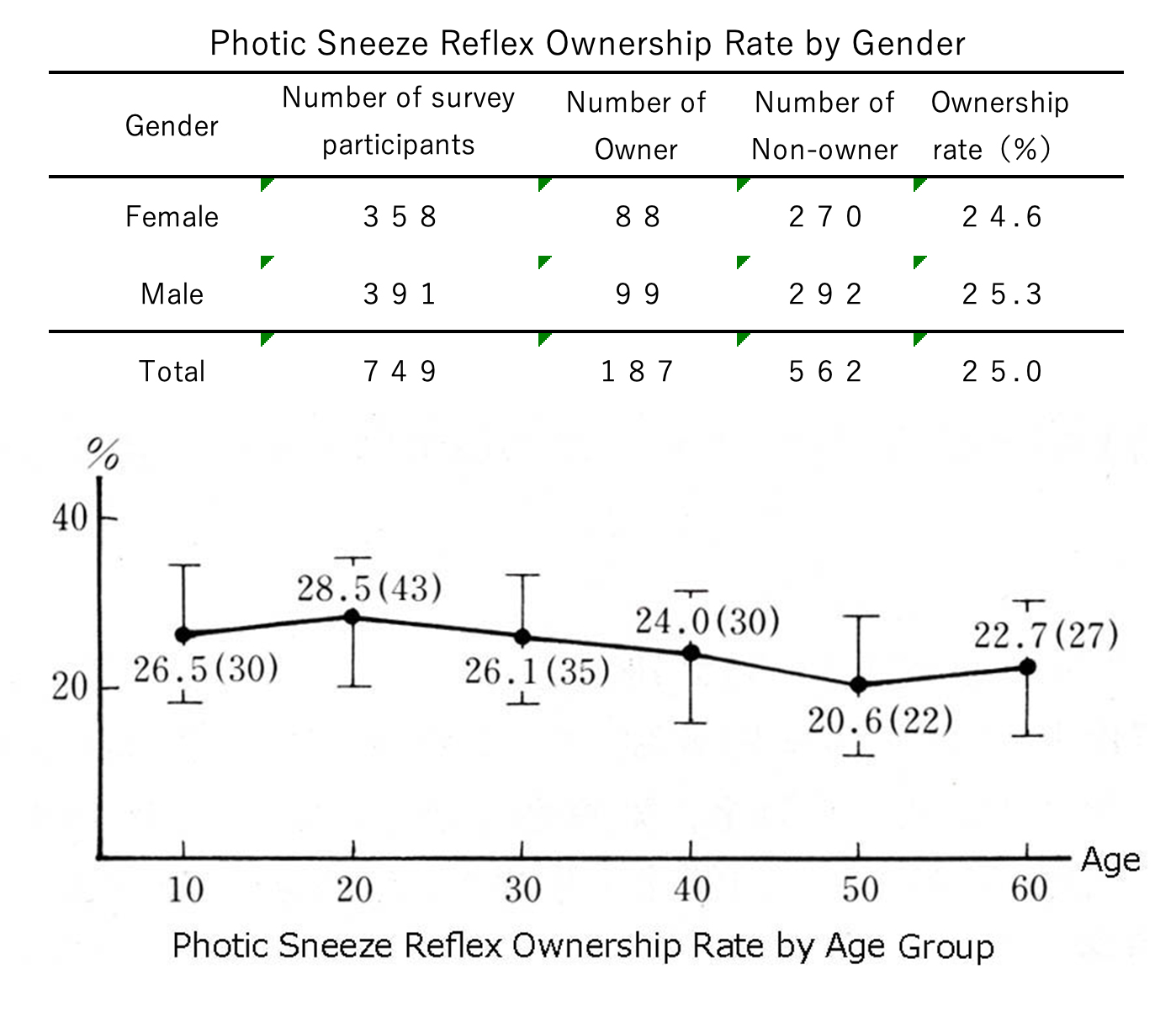
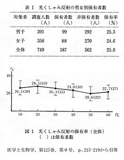 光くしゃみ反射とは、光刺激が誘因となって反射的にくしゃみが起こる現象です。屋内から晴天下の屋外に出た時や太陽の光が直接目に入った時など、まぶしさを感じた瞬間に起こります。光くしゃみ反射は、まぶしさを感じている間だけに起こるので、くしゃみの回数は通常1回か2回で、連続して何回も起こることはありません。光くしゃみ反射は、誰にでも存在するのではなく一部の人たちだけに存在する生理的(正常)な反射です。私たちの749名を対象にした調査において、日本人の約25%の人がこの反射をもっていることがわかりました(医学と生物学、1992年、第125巻 第6号 215-219頁)。しかし、この中には「稀にしか反射が起こらない人」や「若い頃にはよく起こったが今は起こらない」などという人も含まれていますので、実際には光くしゃみ反射が日常的に起こる人はその半分くらいと考えられます。光くしゃみ反射をもつ家系では光くしゃみ反射を持つ人の割合が著しく高いので、光くしゃみ反射は優性遺伝によって子孫に伝えられると推察できます。光くしゃみ反射をもつ人のあいだでもくしゃみを誘発する光の強度には著しい個人差があり、太陽光が直接目に入った時のように強烈な光だけに反射を起こす人もいれば、室内灯の灯り程度の弱光でくしゃみをする人もいます。この現象がどのようなメカニズムによって起こるかについて、私たち研究グループは、以下のように推察しています。
くしゃみは、「鼻腔に入り込んだ異物を鼻汁ごと激しく鼻息で吹き飛ばす反射」です(余分な空気は口から出ます)。鼻腔粘膜が異物の刺激に晒されると、鼻腔粘膜の肥満細胞からヒスタミンが放出され、それにより鼻汁が分泌され、血管も拡張し、鼻にムズムズとした感覚が起こります。そのムズムズした感覚は三叉神経を介して脳の感覚中枢へ、そしてくしゃみ中枢へと伝えられ、くしゃみが誘発されます。くしゃみを引き起こす鼻汁分泌は、鼻腔に異物が入った時だけではなく、脳からの指令が引き金となって起こることもあります。何らかの理由で延髄の唾液核(と呼ばれる神経細胞集団)が興奮すると、唾液核を起始する副交感神経線維(鼻汁分泌と血管拡張指令を伝える神経)は、顔面神経を介して翼口蓋神経節(上顎後深部の翼口蓋窩に存在する神経細胞集団)に到達し、さらに鼻粘膜に神経興奮が伝えられて鼻汁分泌と血管拡張が起こります。すると、鼻粘膜にムズムズした感覚が起こり、これが原因でくしゃみ反射が起こります。
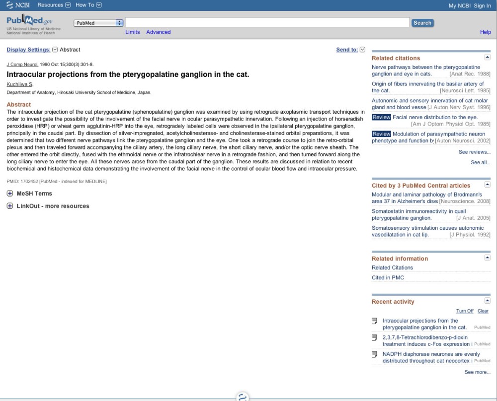
私たち研究グループは、鼻汁を分泌させる翼口蓋神経節は、顔面神経だけではなく、動眼神経系と結合していることを解剖学的に明らかにしました(The Journal of Comparative Neurology, vol. 300: p301-308, 1990)。目に入ったまぶしい光情報は、中脳の視蓋前域オリーブ核に伝えられたあとに動眼神経副核(Edinger-Westphal核:EW核)に伝えられ、さらに眼窩内の毛様体神経節を介して瞳孔括約筋に伝えられ、対光反射(眩しい時に瞳が小さくなる反射)が起こります。私たちは、「EW核を起始した神経軸索を含む動眼神経の小枝(細い神経)が翼口蓋神経節の鼻粘膜支配細胞領域に到達し、EW核は対光反射を起こすのと同時に翼口蓋神経節の鼻汁分泌細胞群と血管平滑筋を刺激する」と推論しています。眩しいと感じる光情報はこの神経路を介して鼻粘膜を刺激し、くしゃみ反射を誘発させると考えられます。
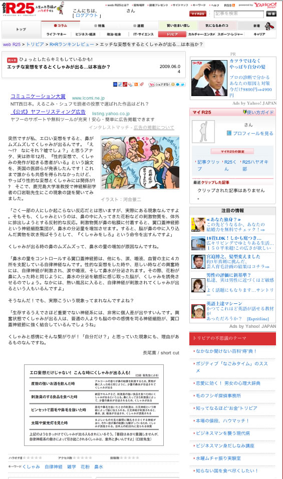
ちなみに、顔面神経は感情に関係する神経系です。顔面神経は、心の動きに伴って顔の表情を作り、涙を流し、顔を紅潮させます。心の激しい動きが鼻汁を分泌させることもありますので、稀ではありますが、エッチなことを考えるとくしゃみが出る人もいます。
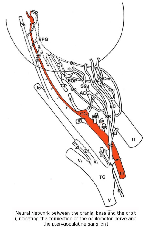
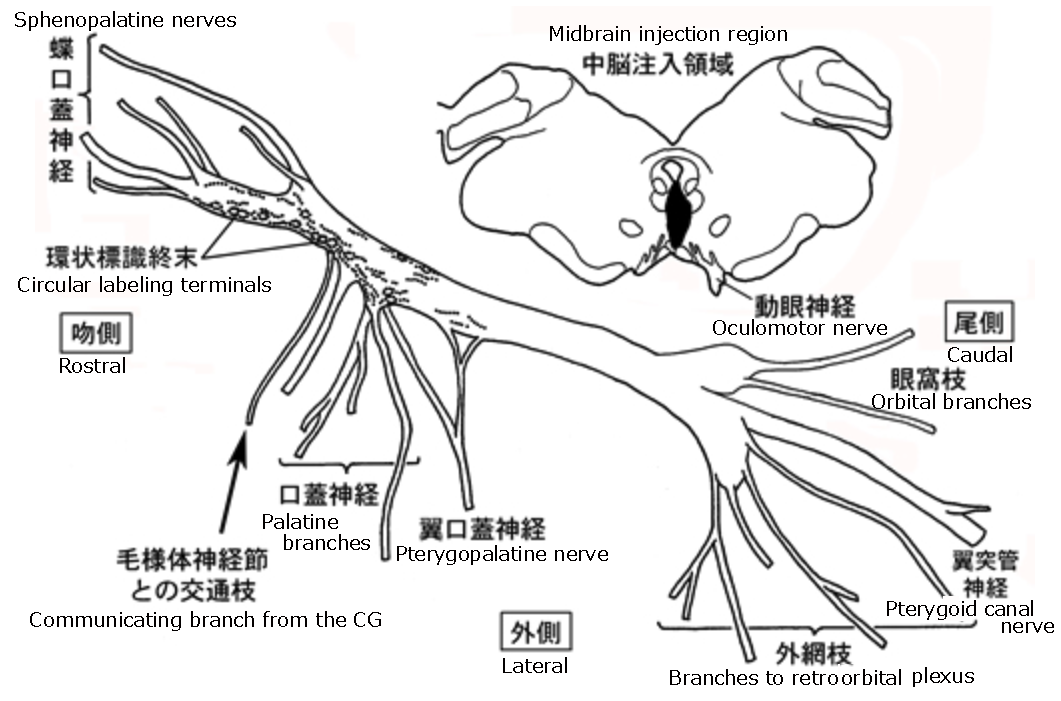
頭蓋底と眼窩の神経網(動眼神経と翼口蓋神経節の結合を示す)
動眼神経副核(EW核)を起始した副交感性節前線維は動眼神経(Ⅲ)に沿って眼窩に入り、毛様体神経節(CG)に達する。毛様体神経節でシナプス形成したあとの節後線維またはこれを素通りした節前線維は、翼口蓋神経節への交通枝(4つの矢頭で示した)を経由し、翼口蓋神経節(PPG)の吻側部に到達する。翼口蓋神経節のこの領域から蝶口蓋神経(Sp)が出て鼻粘膜および口蓋に分布する。Ⅱ、視神経:Ⅴ、三叉神経:ACG、副毛様体神経節:ROP、後眼窩神経叢および神経節。OrとOcは翼口蓋神経節または後眼窩神経節から眼球へ投射する血管調節神経:矢印は動眼神経と三叉神経の交通枝(三叉神経を経由して副毛様体神経節に至る副交感神経節前線維を含む):CBは副毛様体神経節に入る三叉神経根。
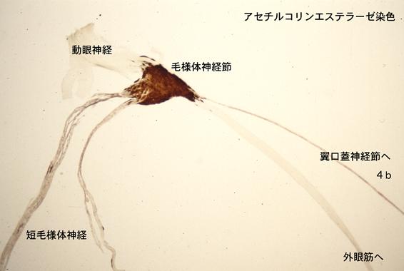
毛様体神経節の顕微鏡写真
動眼神経のアセチルコリンエステラーゼ染色(AChE染色)標本の顕微鏡写真。毛様体神経節から翼口蓋神経節へ向かう神経(4b)には、AChE陽性線維と陰性線維の両方が含まれている。副交感神経の節前線維はAChE陰性であり、節後線維はAChE陽性である。したがって、毛様体神経節から翼口蓋神経節へ向かうAChE陰性線維は、EW核を起始する副交感神経節前線維であり、毛様体神経節を素通りして、翼口蓋神経節の吻側部に存在する鼻粘膜支配細胞に接続する可能性がある。一方、AChE陽性線維は毛様体神経節細胞の節後線維であり、翼口蓋神経節を素通りし、蝶口蓋神経を介して鼻粘膜に分布すると考えられる。これらのことは、動眼神経核が鼻粘膜の血流調節と鼻汁分泌に関わっている可能性を強く示唆している。
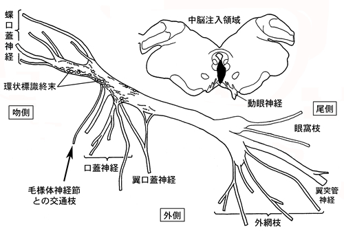
ネコの中脳正中部に標識化合物を微量注射した実験における中脳切片と翼口蓋神経節切片のスケッチ
中脳の注入領域の広がりと翼口蓋神経節における順行性標識の分布を示す。注入領域はEW核(Edinger-Westphal nucleus)を覆っている。翼口蓋神経節吻側部の大型節後ニューロン周囲に環状配列する順行性標識が顕著に認められた。このことは、EW核から翼口蓋神経節吻側部への順行性投射が存在することを示唆している。標識が出現した翼口蓋神経節の吻側部は蝶口蓋神経を介して鼻腔粘膜を自律神経性に支配する。大型細胞は外分泌腺を支配することが知られている。(中外医学社 Clinical Neuroscience, Vol. 33, 2015,「光を見るとくしゃみが出るのはなぜですか?」から引用)
Photic Sneeze Reflex
 The phenomenon of photic sneeze reflex is the occurrence of reflexive sneezing triggered by exposure to light stimuli. It happens at the moment when one experiences brightness, such as when moving from indoors to the outdoor sunshine or when direct sunlight enters the eyes. The photic sneeze reflex occurs only while perceiving brightness, so the number of sneezes is typically one or two, and it does not happen repeatedly.
The photic sneeze reflex is a physiological (normal) reflex that exists only in some individuals, not everyone. In our survey study conducted with a sample of 749 individuals, it was found that approximately 25% of Japanese people exhibit this reflex (Medicine and Biology, 1992, Vol. 125, No. 6, pp. 215-219). However, within this group, there are individuals who rarely experience the reflex or had it frequently in their youth but not anymore. Therefore, it is estimated that only about half of those with the photic sneeze reflex experience it regularly. The photic sneeze reflex is expressed within families, suggesting that it is likely transmitted to descendants through dominant inheritance. Even among individuals with the photic sneeze reflex, there is a significant individual variation in the light intensity that induces sneezing. Some people only experience the reflex with intense light, such as direct sunlight, while others may sneeze with weak light, like indoor lighting. Regarding the mechanism behind this phenomenon, our research group speculates as follows.
Sneezing can occur not only when foreign substances enter the nasal cavity but also in response to signals from the brain. For some reason, when a group of nerve cells called the salivary nucleus in the medulla oblongata becomes excited, parasympathetic nerve fibers originating from it reach the pterygopalatine ganglion (a group of nerve cells located in the pterygopalatine fossa deep behind the upper jaw) through the facial nerve. Furthermore, nerve excitation is transmitted to the nasal mucosa, leading to nasal mucus secretion and blood vessel dilation. As a result, a tingling sensation occurs in the nasal mucosa, which is the cause of the sneezing reflex.
Our research group anatomically demonstrated that the pterygopalatine ganglion is not only controlled by the facial nerve but also by the oculomotor nerve (The Journal of Comparative Neurology, vol. 300: p301-308, 1990). Intense light entering the eyes is transmitted to the pretectal olivary nucleus in the midbrain and further relayed to the Edinger-Westphal nucleus (EW nucleus). The parasympathetic nerves originating from the EW nucleus are conveyed through the ciliary ganglion located behind the eyeball to the pupillary constrictor muscle, causing the pupillary light reflex (contraction of the pupils in response to bright light). We discovered that a portion of the nerves originating from the EW nucleus reaches the nasal mucosa control cell region of the pterygopalatine ganglion. It can be inferred that the EW nucleus, while inducing the pupillary light reflex, simultaneously stimulates the nasal mucosa control cell group in the pterygopalatine ganglion, leading to vasodilation and nasal mucus secretion.
Incidentally, the facial nerve is part of the neural system associated with emotions. The facial nerve is responsible for creating facial expressions, shedding tears, and causing facial flushing in response to emotional movements. Intense emotional states can also lead to nasal mucus secretion and vasodilation, so although rare, some individuals may experience sneezing when indulging in sexual fantasies. (Bhutta and Maxwell, 2008)

The parasympathetic preganglionic fibers originating from the Edinger-Westphal nucleus (EW nucleus) enter the orbit along the oculomotor nerve (III) and reach the ciliary ganglion (CG). A nerve that pass through the ciliary ganglion (indicated by four arrowheads) then travel to reach the rostral part of the pterygopalatine ganglion (PPG). From this region of the ganglion, the sphenopalatine nerve (Sp) emerges, distributing to the nasal mucosa and palate. II, optic nerve; V, trigeminal nerve; ACG, accessory ciliary ganglion; ROP, posterior orbital nerve and ganglion. Or and Oc represent vasomotor nerves projecting to the eyeball from the pterygopalatine ganglion or posterior orbital ganglion. Arrow indicate communicating branches between the trigeminal nerve and the ciliary nerve (including parasympathetic preganglionic fibers reaching the ciliary ganglion via the trigeminal nerve); CB represents the trigeminal root entering the ciliary ganglion.
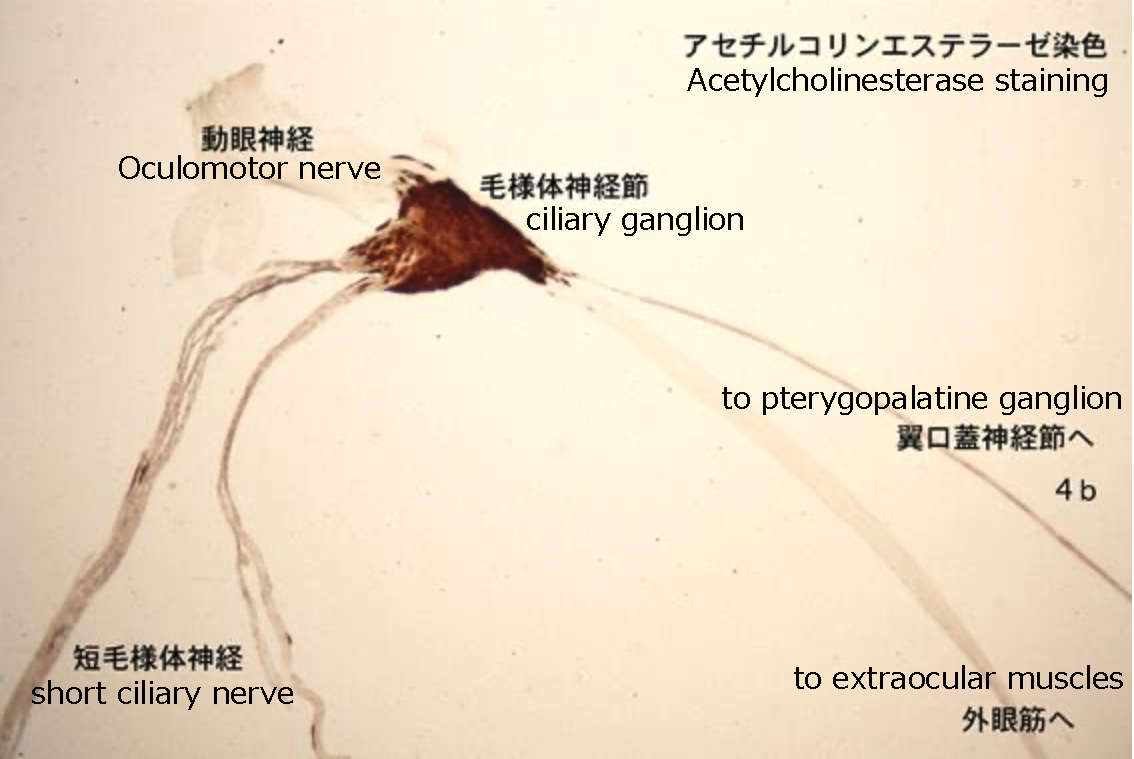
Microscopic photographs of acetylcholinesterase staining (AChE staining) specimens of the oculomotor nerve. In the nerves traveling from the ciliary ganglion to the pterygopalatine ganglion (4b), both AChE-positive and AChE-negative fibers are present. The preganglionic fibers of the parasympathetic nervous system are AChE-negative, while the postganglionic fibers are AChE-positive. Therefore, AChE-negative fibers traveling from the ciliary ganglion (CG) to the pterygopalatine ganglion (PPG) may originate from the Edinger-Westphal (EW) nucleus, serving parasympathetic preganglionic fibers to nasal mucosa control cells located in the rostral part of the PPG. On the other hand, AChE-positive fibers are postganglionic fibers of the CG cells, and they may pass through the PPG, distributing to the nasal mucosa via the sphenopalatine nerve (Sp). These findings strongly suggest the potential involvement of the EW nucleus in the regulation of blood flow and nasal secretion in nasal mucosa.
Midbrain sections and pterygopalatine ganglion sections in an experiment involving microinjections of a tracer substance into the region containing the Edinger-Westphal (EW) nucleus in cats: illustrating the injection site of the tracer substance and showing the distribution of anterograde labeling.
The injection site covers the Edinger-Westphal nucleus (EW nucleus). Anterograde labeling, arranged in a circular pattern around large postganglionic neurons in the rostral part of the pterygopalatine ganglion (PPG), was prominently observed. This suggests the presence of anterograde projections from the EW nucleus to the rostral part of the PPG, where labeling appeared is known to autonomically control the nasal mucosa through the sphenopalatine nerve (Sp). Large cells of the PPG are known to control exocrine glands (quoted from "Why do we sneeze when we see the light?" Clinical Neuroscience, Vol. 33, 2015).
